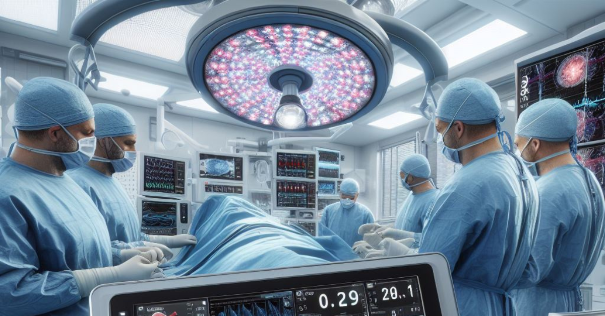
CIPTV is an advanced technological platform that enhances the precision and effectiveness of minimally invasive tumor surgeries through the integration of real-time imaging and computer algorithms. This technology combines various aspects of medical imaging, such as ultrasound, CT scans, and MRI with artificial intelligence and robotic assistance, to provide surgeons with detailed visualizations and guidance during procedures. The importance of CIPTV in modern surgery cannot be overstated; it minimizes surgical risks, enhances accuracy, reduces recovery times, and improves overall surgical outcomes by enabling precise targeting and minimal disruption to surrounding healthy tissues. As CIPTV systems incorporate increasingly sophisticated machine learning algorithms, they not only improve the capabilities of current surgical practices but also promise to revolutionize patient care by making surgeries safer, quicker, and more efficient.
The Basics of CIPTV
What is CIPTV?
CIPTV, which stands for Computer-Integrated Percutaneous Tumor Visualization, is a cutting-edge medical technology that revolutionizes the way surgeries, particularly tumor removals, are performed. This technology combines real-time imaging, computer algorithms, and advanced surgical tools to enable precise and minimally invasive interventions. The role of CIPTV in surgery is crucial as it enhances the surgeon’s ability to accurately target and remove tumors with minimal impact on surrounding healthy tissues, thus improving patient outcomes, reducing recovery times, and decreasing the risks associated with more invasive procedures. Key components of CIPTV include high-resolution imaging technologies like MRI and CT scans, data processing software that provides real-time feedback and visualization during surgery, and robotic systems that assist with precise movements and manipulations. Together, these technologies ensure that CIPTV is at the forefront of modern surgical practices, offering a blend of accuracy, efficiency, and safety previously unattainable in complex surgical scenarios.
Historical Perspective
The historical perspective of neurophysiological monitoring and the development of Computer-Integrated Percutaneous Tumor Visualization (CIPTV) reflect significant milestones in medical technology, showing a robust evolution over the decades. Initially, neurophysiological monitoring began in the early 20th century with basic EEGs used during surgical procedures to monitor the brain’s electrical activity. This form of monitoring has advanced dramatically, incorporating complex modalities like motor evoked potentials and somatosensory evoked potentials, which help surgeons avoid vital nerve functions during operations. In parallel, the development of CIPTV marked a revolutionary step in the late 20th and early 21st centuries, evolving from simple image-guided surgical techniques to sophisticated systems integrating real-time imaging, 3D visualization, and robotic assistance. Key milestones in CIPTV include the integration of MRI and CT technologies with live surgical navigation systems in the 1990s, followed by the introduction of AI algorithms for improved imaging analysis and surgical precision in the 2000s. These advancements have collectively enhanced the capability of surgeons to perform precise, minimally invasive procedures, thereby improving patient outcomes and setting new standards in surgical care.
CIPTV Technologies
Monitoring Techniques
In the realm of CIPTV technologies, advanced monitoring techniques play a critical role in enhancing the safety and efficacy of surgical procedures. Among these, Motor Evoked Potentials (MEP) and Somatosensory Evoked Potentials (SSEP) are paramount for ensuring the integrity of neural pathways during surgery. MEPs are used to monitor the functionality of motor pathways by delivering small electrical impulses through the scalp to induce a motor response in muscles, which is particularly crucial during surgeries that risk affecting the spinal cord or motor cortex. SSEPs, on the other hand, measure the response of the sensory pathways to similar electrical stimuli. These stimuli are applied to peripheral nerves, and the resultant responses are recorded from the cerebral cortex. The feedback provided by MEPs and SSEPs allows surgeons to continuously assess and safeguard neurological function in real time, significantly reducing the risk of postoperative deficits. This continuous feedback is integral to the CIPTV framework, as it ensures that tumor removal or other targeted surgical actions do not compromise the patient’s neurological health, thereby optimizing surgical outcomes while preserving vital functions.
Advanced Imaging and Integration
Advanced imaging and integration are cornerstone technologies in the framework of Computer-Integrated Percutaneous Tumor Visualization (CIPTV), providing crucial insights that guide surgical precision and decision-making. Within this context, MRI (Magnetic Resonance Imaging) and CT (Computed Tomography) scans are pivotal. They offer detailed cross-sectional views of soft tissue and bony structures, respectively, allowing for precise planning and execution of minimally invasive surgeries. The role of MRI in CIPTV is particularly significant for its superior soft tissue contrast, which is ideal for identifying and delineating tumors in complex anatomical regions. CT scans complement this by providing excellent spatial resolution that is critical in navigating bony environments.
Furthermore, CIPTV harnesses these imaging modalities through real-time data integration and visualization. This integration enables the dynamic overlay of live surgical views with pre-operative imaging data, creating a comprehensive visual map that surgeons can refer to during procedures. This real-time visualization not only enhances accuracy but also adapts to changes during surgery, such as shifts in patient positioning or changes in the target area due to surgical manipulations. This capability is facilitated by sophisticated software and hardware that process and display imaging data instantaneously, thereby supporting surgeons in making informed decisions rapidly and maintaining high precision throughout the surgical process. These advancements underscore the transformative impact of modern imaging and real-time data integration in enhancing the efficacy and safety of surgical interventions through CIPTV technologies.
Applications of CIPTV
Orthopedic Surgery
In orthopedic surgery, particularly in procedures involving the spinal cord, the application of Computer-Integrated Percutaneous Tumor Visualization (CIPTV) has become instrumental in enhancing surgical precision and patient safety. CIPTV utilizes advanced imaging and real-time data integration to provide detailed visualizations, enabling surgeons to navigate complex spinal structures carefully. Critical to this process is the use of neurophysiological monitoring techniques like Motor Evoked Potentials (MEPs) and Somatosensory Evoked Potentials (SSEPs), which continuously assess the functional integrity of the spinal cord and nerve pathways during surgery. This capability is essential for preventing nerve damage; it allows surgeons to detect potential impairments and adjust their techniques promptly, thus minimizing the risk of postoperative neurological deficits and ensuring better overall surgical outcomes while preserving vital motor and sensory functions.
Neurosurgery
In neurosurgery, Computer-Integrated Percutaneous Tumor Visualization (CIPTV) significantly enhances the precision and safety of brain surgeries and tumor resections. By integrating advanced imaging technologies such as MRI and CT scans with real-time data visualization, CIPTV allows neurosurgeons to meticulously plan and execute procedures, minimizing damage to critical brain structures. This precision is particularly crucial in the delicate task of removing brain tumors. Additionally, CIPTV proves invaluable in cerebral aneurysm procedures, where real-time imaging and visualization assist surgeons in accurately positioning clips or navigating endovascular devices to treat aneurysms with techniques like coiling or stenting. The capability to monitor vascular changes instantly during these procedures reduces the risk of complications such as bleeding or secondary stroke, thus significantly improving patient outcomes in complex neurosurgical interventions.
Cardiothoracic Surgery
In cardiothoracic surgery, Computer-Integrated Percutaneous Tumor Visualization (CIPTV) is transforming the approach to both cardiac and thoracic procedures by enhancing precision and safety through advanced monitoring capabilities. During cardiac surgeries, CIPTV’s integration of real-time imaging and data visualization allows for meticulous monitoring of the heart’s function and structure, facilitating delicate interventions such as valve replacements or repairs and coronary bypass surgeries with reduced risk to the patient. In thoracic procedures, particularly those involving tumor resections from the lungs or other chest structures, CIPTV aids surgeons by providing detailed 3D images and real-time feedback. This capability ensures that surgical maneuvers are precisely targeted, minimizing damage to surrounding vital organs and tissues. By improving the precision of surgical techniques and enhancing the surgeon’s ability to respond to intraoperative changes, CIPTV significantly betters patient outcomes in the realm of cardiothoracic surgery.


